
Prostate Cancer SCreening
Cancer screening means looking for cancer before it causes symptoms. The goal of screening for prostate cancer is to find cancers that may be at high risk for spreading if not treated, and to find them early before they spread.
The initiation and frequency for prostate cancer screening depends on a variety of risk factors including family history of cancer, ethnicity, results of genetic testing, previous PSA levels, and more. A doctor at the Prostate Cancer Center of Excellence can help you put together an appropriate screening plan that may include digital rectal exams, PSA tests, and diagnostic imaging.
If you have prostate cancer, you are not alone – approximately 220,000 men are diagnosed in the United States each year. Other than skin cancer, prostate cancer is the most commonly diagnosed cancer among men. However, it is also one of the most treatable, especially if detected early.

Prostate-specific antigen (PSA) test
Prostate-specific antigen (PSA) is a protein produced by the prostate gland. The PSA test is a blood test that measures the level of PSA in a man’s blood.
The PSA test is commonly used as a screening test for prostate cancer, as elevated levels of PSA can be a sign of prostate cancer. However, PSA levels can also be elevated due to non-cancerous conditions such as benign prostatic hyperplasia (BPH) or inflammation of the prostate gland (prostatitis), as well as age.
If PSA levels are elevated or if other symptoms or signs of prostate cancer are present, a doctor may recommend additional tests, such as a digital rectal exam or a biopsy, to confirm the diagnosis of prostate cancer.
Men with very low PSA levels may need to be tested every two years. If PSA is higher, the doctor may recommend more frequent testing.
PSA tests can also be used in men who have been diagnosed with prostate cancer. For instance, they may:
- Help doctors plan treatment or further testing
- Determine if cancer has metastasized (spread beyond the prostate)
- Find out if treatment is working or cancer has returned
- Aid in active surveillance (also called watchful waiting) by showing if cancer is growing

Digital Rectal Exam (DRE)
A digital rectal exam (DRE) is a physical exam performed by a doctor to check the prostate gland for any abnormalities, including signs of prostate cancer. During a DRE, the doctor inserts a gloved, lubricated finger into the rectum and feels the prostate gland for any lumps, bumps, or hard areas that may be indicative of cancer.
DREs are a quick and simple exam that can be performed in a doctor’s office. It may be done in conjunction with other tests, such as a prostate-specific antigen (PSA) blood test, to help diagnose prostate cancer. However, it is important to note that not all prostate cancers can be detected by a DRE, and false positives and false negatives can occur.
The DRE is not a definitive cancer test, but regular exams help detect changes in the prostate over time. These changes might signal cancer or pre-cancerous conditions.
Imaging Exams
Imaging exams are often used to detect and stage prostate cancer, as well as to monitor the disease and assess the effectiveness of treatment. Some imaging exams that may be used for prostate cancer include:
- Transrectal ultrasound: During a transrectal ultrasound, a small probe is inserted through the rectum. This probe generates high-energy sound waves that bounce off your tissue and create a picture of your prostate gland on a screen. This picture can be examined for abnormalities or tumors.
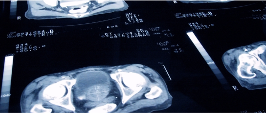
- Computed tomography (CT) scan: CT scans use X-rays to create detailed cross-sectional images of the body. CT scans can help detect whether prostate cancer has spread to nearby lymph nodes or other organs, such as the bones.
- Positron emission tomography (PET) scan: PET scans use a small amount of radioactive material to create images of the body’s cells and tissues. PET scans can be used to detect whether prostate cancer has spread beyond the prostate gland, including to distant organs such as the bones.
- Magnetic resonance imaging (MRI): MRI uses a magnetic field and radio waves to create detailed images of the prostate gland and surrounding tissues. MRI can help determine the size and location of a tumor, as well as whether the cancer has spread to nearby lymph nodes or other organs.
The choice of imaging test will depend on a variety of factors, including the individual’s specific case and the stage of their prostate cancer. It is important to note that while imaging exams can be helpful in diagnosing and monitoring prostate cancer, they may not always detect small tumors or distinguish between cancerous and non-cancerous lesions.

Biopsy
If one of these screening tests above indicates that there might be cancer present, your doctor may order a biposy.